- Foot and Ankle Anatomy
- Foot and Ankle Conditions
- Foot and Ankle Procedures
Plantar Fasciitis

Plantar fasciitis refers to the inflammation of the plantar fascia, a thick band of tissue that is present at the bottom of the foot. It runs from the heel bone to the toes and forms the arch of your foot. Plantar fasciitis is one of the most common causes of heel pain. It is most often seen in middle-aged men and women, but may also occur in those who are constantly on their feet.
Ankle Fractures

Ankle injuries are very common in athletes and individuals performing physical work; often resulting in severe pain and impaired mobility. Pain after ankle injuries can either be from a torn ligament (ankle sprain) or broken bone (ankle fracture). An ankle fracture is a painful condition where there is a break in one or more bones forming the ankle joint.
Morton's Neuroma

Morton’s neuroma refers to a nerve injury that occurs between the toes, usually the third and fourth toes. This causes pain and thickening of the nerve tissue. Compression or chronic irritation of this interdigital nerve is the main cause of Morton’s neuroma. Excess pressure exerted on the nerves due to the narrowing of the gap between...
Foot and Ankle Arthritis

Arthritis is the inflammation of joints as a result of degeneration of the smooth cartilage that lines the ends of bones in a joint. This degeneration of the cartilages leads to painful rubbing of the bones, swelling, and stiffness in the joints, resulting in restricted movements. Arthritis in the foot and ankle can occur due to fractures, dislocation, inflammatory disease, or congenital deformity.
Ankle Rheumatoid Arthritis

Arthritis is inflammation in a joint as a result of cartilage degeneration causing joint pain, swelling, stiffness, and restricted movement. Arthritis of the foot and ankle joint can occur due to fractures, dislocation, inflammatory disease, or congenital deformity. Rheumatoid arthritis is an auto-immune disease in which the body’s immune system...
Foot Rheumatoid Arthritis

Arthritis is inflammation in the joint resulting from the degeneration of cartilage causing joint pain, swelling, and stiffness resulting in restricted movements. Arthritis of the foot and ankle joint can occur due to fractures, dislocation, inflammatory disease, or congenital deformity.
Midfoot Arthritis

Midfoot arthritis is pain and inflammation of the midfoot. It occurs due to damage of cartilage or tissues around the joints. The damage may occur due to injury, aging or autoimmunity. The foot bones are the phalanges, the metaphalanges, and the tarsal bones. The midfoot consists of 5 bones called lesser tarsal bones.
Hallux Rigiditis

Hallux Rigiditis is a form of degenerative arthritis at the metatarsophalangeal or MTP joint where the base of your big toe attaches to the foot. Arthritis is the inflammation of joints as a result of degeneration of the smooth cartilage that lines the ends of bones in a joint.
Bunion
A bunion, also known as hallux valgus, is a bony protuberance that appears on the outer surface of the big toe when it angles toward the adjacent toe. It is an extra bone and a fluid-filled sac that grows at the base of the big toe. Bunions are common in women and tend to run in families (heredity).
Ankle Pain

Ankle pain refers to any form of pain or discomfort affecting the ankle joint. Your ankles are more prone to injury and pain since they bear the weight of your entire body. Your doctor will review your symptoms and medical history and a physical examination of the ankle will be conducted.
Ankle Sprain

A sprain is the stretching or tearing of ligaments. Ligaments connect adjacent bones and provide stability to a joint. An ankle sprain is a common injury that occurs when you suddenly fall or twist the ankle joint, or when you land your foot in an awkward position after a jump. Most commonly, it occurs when you participate in sports, or jump or run on a surface that is irregular.
Ankle Instability

The joints of the ankle are held in place and stabilized by strong bands of tissue called ligaments. Ankle instability is a chronic condition characterized by a recurrent slipping of the outer side of the ankle. It usually results from repeated ankle sprains, which are injuries to the ligaments. Ankle instability is generally noticed when you move your ankle joint but can also occur while standing.
Foot Fracture

Trauma and repeated stress can cause fractures in the foot. Extreme force is required to fracture the bones in the hindfoot. The most common type of foot fracture is a stress fracture that occurs when repeated activities produce small cracks in the bones. The hindfoot is separated from the midfoot by the mediotarsal joint and the midfoot is separated from the forefoot by the Lisfranc joint.
Ankle Ligament Injury

An ankle ligament injury, also known as an ankle sprain, can be caused by a sudden twisting movement of the foot during any athletic event or during daily activities. When stretched beyond its limit, the ligament may partially or completely tear. The injury can range from mild to severe, depending on the condition of the injured ligament and the number of ligaments involved.
Foot and Ankle Trauma
Foot and ankle trauma refers to injuries that affect the bones, joints, ligaments, tendons, or soft tissues of the foot and ankle. These injuries can result from accidents, falls, sports activities, poor training practices, high-energy trauma, such as motor vehicle accidents, or from the use of improper gear.
Ankle Dislocation

Ankle dislocation is a condition that occurs when the bones of the lower leg called the fibula and tibia get separated from the talus or ankle bone. This can cause serious damage to the nerves and ligaments surrounding the ankle, leading to a decline in strength and overall health of the leg.
Achilles Tendonitis

Inflammation of the Achilles tendon is known as Achilles tendonitis or tendinitis. The Achilles tendon is a tough band of fibrous tissue that runs down the back of your lower leg and connects your calf muscle to your heel bone. The tendon is used when you walk, climb, jump, run and stand on your tip toes.
Ankle Impingement

The ankle is made up of many bones, muscles, and ligaments that aid in the smooth movement of the foot. Ankle impingement is the painful limitation of movement of the ankle due to an abnormality in the soft tissue or bone. Impingement can occur in the ankle's front (anterior ankle impingement) or back (posterior ankle impingement).
Achilles Tendon Bursitis

Achilles tendon bursitis or retrocalcaneal bursitis is a condition that commonly occurs in athletes. It is a painful condition caused by the swelling of the bursa, a fluid-filled sac that is located at the back of the heel under the Achilles tendon. This retrocalcaneal bursa contains a lubricating fluid that acts as a cushion to reduce friction between muscle and bones.
Bunionette

Bunionette also referred to as a tailor’s bunion is a bony lump that grows on the outside of the foot at the base of your little toe. The deformity got its name as q tailor’s bunion when tailors once sat with their legs crossed all day, with the outside edge of their feet rubbing on the ground.
Foot Problems

The foot is a highly complex part of the human body that has an integral role in most activities of daily living. It consists of 26 bones connected with joints, ligaments, and tendons. It is susceptible to injury due to significant stress caused by supporting the weight of the entire body when walking or running.
Toe Fractures

A fracture is defined as a break in the continuity of the bone when a force against your body is too strong for the bone to bear. A toe fracture is when one or more of the toe bones of the foot are broken after severe trauma to the toes or foot. Toe fractures are commonly divided into 2 types namely, traumatic fracture and stress fracture.
Cavus Foot Deformity (High Arches)
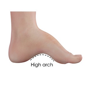
Cavus foot also referred to as a high arch, is a condition in which the arch on the bottom of the foot that runs from the toes to the heel is arched more than normal. Because of this, excessive weight falls on the ball and heel of the foot when walking or standing, causing pain and instability.
Lesser Toe Deformities

Lesser toe deformity is an abnormality in the anatomy of your toe that occurs as a result of imbalance between the intrinsic and extrinsic muscles. Lesser toes in your foot are those other than the big toes and together stabilize your foot while standing and help in balancing the body.
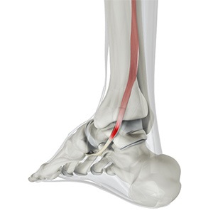
Achilles Tendon Rupture

The Achilles tendon is a strong fibrous cord present behind the ankle that connects the calf muscles to the heel bone. It is used when you walk, run and jump. The Achilles tendon ruptures most often in athletes participating in sports that involve running, pivoting and jumping. Recreational sports that may cause Achilles rupture include tennis, football, basketball, and gymnastics.
Diabetic Foot Conditions

Diabetes is a chronic condition that is characterized by high blood glucose (sugar) levels. Diabetic patients are at high risk for developing chronic wounds, especially in the feet. If left untreated, these wounds can cause serious problems that can lead to infections and eventually gangrene, which may require amputation.
Flatfoot

Flatfoot, also known as “fallen arches” or Pes planus, is a deformity in children’s feet where the arch that runs along the sole of the foot collapses to the ground or is not formed at all. Flatfoot is normal in the first few years of life as the arch of the foot usually develops between the age of 3 and 5 years.
Ankle Tendonitis

Ankle tendonitis is an inflammation of a tendon located in your ankle. Tendonitis can usually happen due to overuse of your ankle with repetitive movements or stretching. The ankle is one of the most common areas to develop tendonitis over other parts of the body. It is also sometimes referred to as ankle tendinitis.
Hammertoe

A hammertoe is a deformity of a lesser toe (second through fifth toes), where the toe is bent upward at the toe’s middle joint, resembling a hammer. The bent portion may rub against the shoe causing pain, irritation and develop corns. This condition is caused by wearing shoes that are too tight or narrow near the toes...
Hallux Limitus

Hallux limitus is a condition characterized by stiffness and decreased movement of the big toe. As the condition progresses, bone spurs and arthritis may develop in the toe joint (metatarsophalangeal joint), eventually leading to complete joint rigidity.
Lisfranc (Midfoot) Fracture
The Lisfranc joint or tarsometatarsal joint refers to the region in the middle of the foot. It is a junction between the tarsal bones (bones in the foot arch) and metatarsal bones (five long bones in the foot). Lisfranc fractures can occur due to a fall from a height or a traumatic motor vehicle accident.
High Ankle Sprain

A high ankle sprain, also known as a syndesmotic sprain, is a specific type of ankle injury that affects the ligaments connecting the tibia and fibula bones in the lower leg. This condition is characterized by pain, swelling, and limited mobility in the ankle joint. Unlike traditional ankle sprains, high ankle sprains...
Foot Deformities

Foot deformities are structural abnormalities or misalignments in the bones, muscles, or ligaments of the foot. These deformities can be congenital (present at birth) or acquired due to various factors such as injury, disease, poor footwear, or biomechanical imbalances.
Foot Spasms

Foot spasms, also known as foot cramps, occur due to the involuntary contraction of the foot muscles. These cramps are usually harmless and will get better on their own; however, sometimes they may need medical attention. Based on the intensity of the spasm, the sensation can vary from a tiny prick to an extremely painful condition.
Ankle Contracture

Ankle contracture is a condition characterized by a permanent shortening or tightening of the muscles, tendons, or ligaments around the ankle joint. This can lead to a restricted range of motion and difficulty in moving the ankle. Ankle contracture can significantly impact mobility and quality of life, so early intervention and consistent management are crucial for optimal outcomes.
Stress Fractures of Foot and Ankle

A stress fracture is described as a small crack in the bone which occurs from an overuse injury of a bone. It commonly develops in the weight-bearing bones of the lower leg and foot. When the muscles of the foot are overworked or stressed, they are unable to absorb the stress and when this happens the muscles transfer the stress to the bone which results in stress fracture.
Charcot Foot Deformity

Charcot foot deformity, also known as Charcot arthropathy, is a condition characterized by weakness and inflammation of the bones, joints, and tissues of the foot. If not treated appropriately, it can lead to joint collapse and loss of mobility. Charcot foot deformity is caused by nerve damage that results in loss of sensation in the foot.
Posterior Tibial Tendon Dysfunction
The posterior tibial tendon passes through the ankle to attach the calf muscle with the bones of the midfoot. It provides stability to the arch and supports the foot while walking. Inflammation or a tear of this tendon as a result of injury may cause dysfunction, leading to pain and the development of a flatfoot.
Stiff Big Toe (Hallux Rigidus)

A stiff big toe, also called hallux rigidus, is a form of degenerative arthritis affecting the joint where the big toe (hallux) attaches to the foot. The toe typically becomes stiff at the base and is sometimes called a “frozen joint”. Degenerative arthritis is a medical condition characterized by the chronic breakdown of cartilage in the joints leading to painful inflammation and stiffness.
Claw Toe

Claw toe is a deformity where a toe bends and appears like a bird’s claw. The affected toe is bent upward from the joint at the ball of the foot, and downward at the joints in the middle and tip of the toe to curl under the foot. Hard, thick skin called corns may develop under the ball of the foot or on the top of the affected toe, causing pain while walking.
Turf Toe

Turf toe is an injury to the ligament at the base of the big toe. It is a painful condition that usually results from jamming the toe into the ground or excessive backward bending of the toe. As it is more common in athletes playing on artificial turf, especially those involved in field sports such as football, baseball, and soccer, it is known as turf toe.
Plantar Plate Injuries

Plantar plate injuries refer to damage sustained by one of the strong ligaments located on the bottom of the foot called the plantar plate. The plantar plate is a thick ligament that runs along the ball of the foot, attaching to the joints. It is a fibrocartilaginous structure designed to safeguard the metatarsal head (the long bones of the foot)...
Sesamoid Fracture

A sesamoid fracture is a break in the sesamoid bone. Sesamoids are two small, pea-shaped bones located in the ball beneath the big toe joint at the bottom of the foot. Sesamoid bones are connected to muscles and other bones by tendons that envelop these bones. Sesamoids help the big toe move normally and absorb the weight placed on the ball.
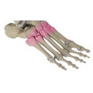
Metatarsal and Phalangeal (Forefoot) Fractures

The forefoot is the anterior or front portion of the foot that functions in weight-bearing and maintaining balance while standing, walking or running. It is formed by 5 metatarsal bones, 14 phalange bones, and various soft tissues. A forefoot fracture is a crack or breaks in any of these bones. In some cases, the broken bone can protrude through the skin. These are called open fractures.
Navicular Stress Fracture

A navicular stress fracture is described as a small crack in the navicular bone (a boat-shaped bone located at the top of the middle part of the foot), which occurs from too much stress being placed on the bone from repetitive activities. Your doctor will review your symptoms, and medical history and perform a thorough physical examination...
Tarsal Coalition

Tarsal coalition is a foot disorder that occurs due to an abnormal connection between two or more tarsal bones. These bones are located at the back of the foot and in the heel. Basically, with this condition, the bones tend to grow within one another and are joined together with the help of surrounding bones and cartilage.
Midfoot Bursitis

Midfoot bursitis is an inflammatory condition affecting the bursae in the middle of the foot. Bursae are small, fluid-filled sacs that reduce friction between bones, tendons, and ligaments. When these sacs become inflamed, it results in pain, swelling, and discomfort in the midfoot region.
Calcaneal Fracture

Calcaneal fractures can be quite severe. Treatment normally includes surgery to reconstruct the normal heel anatomy and restore mobility so that patients can resume their normal activities. However, even after appropriate treatment, some fractures may cause long-term complications, including swelling, pain, loss of movement, and arthritis.
Haglund’s Deformity

Haglund’s deformity, also known as “pump bump”, is a condition in which bony extensions are formed at the back of the heel, leading to swelling near the Achilles tendon. Due to the bony enlargement, the tissue near the Achilles tendon becomes irritated when it rubs against footwear, causing painful bursitis.
Sever’s Disease

Sever’s disease is a painful inflammation of the growth plate in the heel. Growth plates are areas at the end of children’s bones that undergo changes so bone growth can occur. During this time, the muscles and tendons may not grow as fast as the bone causing tightness and pressure at the back of the heel.
Tarsal Tunnel Syndrome

The tarsal tunnel is a narrow passageway that lies on the inside of your ankle and runs into the foot. It encloses arteries, veins, tendons and nerves that supply the foot. The tunnel holds very limited space as it is formed between bones and overlying fibrous tissues. Within the tarsal tunnel lies and runs a nerve called ‘posterior tibial nerve’.
Peroneal Tendonitis

Peroneal tendonitis is inflammation of the peroneal tendons in the ankle due to an acute or overuse injury. It is common in those participating in sports involving repeated ankle movement such as running. The peroneal tendons are two tendons running on the outside of the ankle. They help stabilize the ankle to prevent twisting or sprain injuries.
Sinus Tarsi Syndrome

Sinus Tarsi syndrome(STS) is a painful condition that occurs as a result of trauma or injury to the ligaments in the sinus tarsi area of the foot. The sinus tarsi is a tunnel formed by the talus and the calcaneus bones located in the outer aspect of the hindfoot slightly below and towards the front of the ankle joint.
Osteochondral Lesions of the Ankle

The tibia and the fibula bones of the lower leg join with the talus bone to form the ankle joint. The talus bone is an important bone located between the tibia and fibula and the heel bone (calcaneus). OCL or OCD is the damage to the cartilage and the talus bone of the ankle joint. Usually, the inner or the medial portion of the ankle is affected.